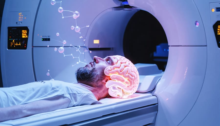
Picture yourself walking into a doctor’s office twenty years ago and asking to see what cannabis does to your brain in real-time. You’d likely have been shown the door. Today, sophisticated brain imaging technology has transformed this impossibility into cutting-edge neuroscience, revealing precisely how Delta-9 tetrahydrocannabinol—the primary psychoactive compound in cannabis—interacts with the intricate landscape of our neural circuitry.
Delta-9 THC neuroimaging represents a fascinating convergence of ancient plant medicine and modern technology. Using tools like functional MRI scanners, PET imaging, and electroencephalography, researchers can now watch the human brain respond to THC molecule by molecule, region by region. These studies aren’t just academic exercises—they’re answering questions that matter deeply to millions: Why does cannabis affect memory? What happens during a “high” at the neurological level? Can we predict who might experience adverse reactions?
The findings have been revelatory, sometimes confirming age-old intuitions about cannabis, other times overturning assumptions we’ve held for decades. We’ve discovered that THC doesn’t simply “turn off” brain regions or dull cognitive function uniformly. Instead, it orchestrates a complex symphony of changes—quieting some neural networks while amplifying others, disrupting communication between certain brain areas while strengthening connections elsewhere.
This technology has given us an unprecedented window into one of humanity’s oldest relationships with a psychoactive substance. The images emerging from these studies tell stories about reward circuits, memory formation, emotional processing, and consciousness itself—stories that are reshaping our understanding of both cannabis and the brain.
Delta-9 THC: The Molecule Behind the Experience
When someone mentions cannabis, they’re usually talking about the effects of a single molecule: Delta-9-tetrahydrocannabinol, or Delta-9 THC for short. This compound is the primary psychoactive ingredient in marijuana, the one responsible for that characteristic “high” that has made cannabis both celebrated and controversial throughout human history.
Think of the cannabis plant as a chemical factory producing over 100 different cannabinoids—molecular cousins that each interact with our bodies in unique ways. Delta-9 THC stands out from its relatives like CBD (cannabidiol) because of one crucial difference: it fits perfectly into specific receptors in our brains, much like a key sliding into a lock. CBD, by contrast, is more like a key that doesn’t quite fit the same locks, which explains why it doesn’t produce intoxicating effects.
To understand how Delta-9 THC works, we need to talk about a remarkable discovery from the 1990s: our bodies actually produce their own cannabis-like chemicals. Scientists call this the endocannabinoid system, and it’s essentially your brain’s own internal messaging network. Imagine your nervous system as a vast city with billions of communication hubs. At these hubs, chemical messengers jump from one nerve cell to another, carrying information about pain, pleasure, memory, and mood.
The endocannabinoid system works like a feedback loop at these communication points. Your body naturally produces molecules called endocannabinoids—think of them as traffic controllers that help regulate the flow of signals between neurons. They bind to specialized receivers called CB1 receptors, which are extraordinarily abundant in the brain, particularly in areas controlling movement, memory, pleasure, and concentration.
Here’s where Delta-9 THC enters the picture: its molecular structure mimics our natural endocannabinoids so closely that it can activate these same CB1 receptors. But unlike our body’s carefully calibrated internal chemicals, THC floods the system like an imposter with an all-access pass, triggering these receptors far more powerfully and persistently than nature intended. This molecular hijacking is what creates the altered perceptions, mood changes, and cognitive effects that neuroimaging studies have now made visible for the first time.
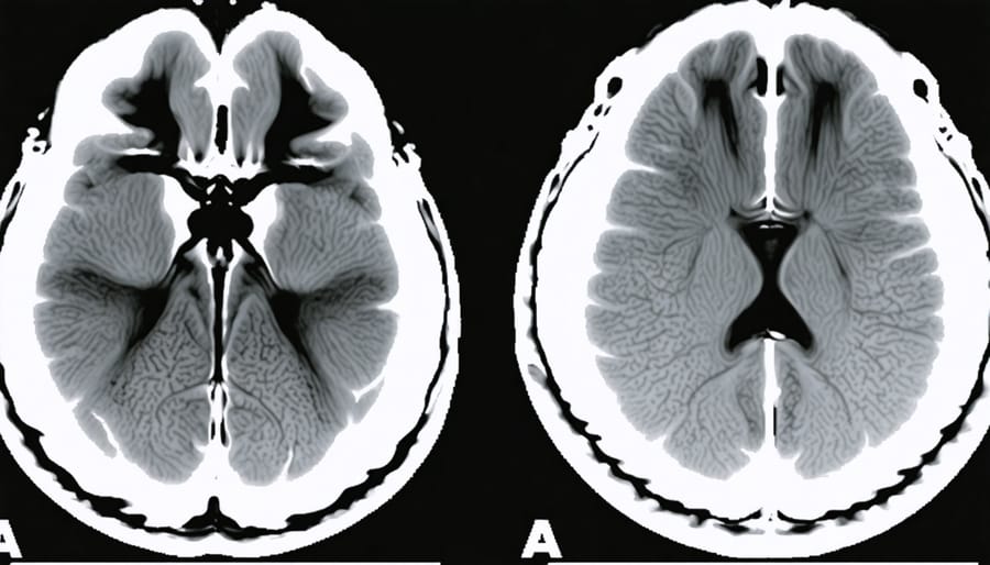
Modern neuroimaging technology like fMRI allows researchers to observe brain activity changes in real-time during THC studies.
Reading the Brain: How Scientists Watch THC in Action
fMRI: Watching Blood Flow Tell the Story
Think of your brain as a bustling city at rush hour. When you perform different tasks—remembering a song, solving a puzzle, feeling anxious—various neighborhoods light up with activity. Functional MRI, or fMRI, works like a traffic helicopter hovering above, monitoring which areas experience increased blood flow. More blood means more oxygen, which means those brain regions are working harder.
Here’s the beautiful simplicity: active neurons are hungry neurons. When a brain region springs into action, blood rushes in to deliver oxygen and glucose, much like food trucks converging on a construction site during lunch break. The fMRI scanner detects these changes in blood oxygen levels, creating colorful maps that show which brain neighborhoods are buzzing with activity.
When researchers study THC’s effects using fMRI, they’re essentially watching the city’s traffic patterns shift in real-time. They might scan someone’s brain before THC consumption, then again afterward, comparing the “before” and “after” maps. These studies have revealed something fascinating: THC doesn’t just turn brain regions “on” or “off” like light switches. Instead, it modulates activity—sometimes dampening overly active areas, sometimes enhancing communication between regions that don’t normally chat much. Similar to other brain stimulation research, these imaging techniques help scientists understand how external substances influence our neural networks in ways we’re only beginning to comprehend.
PET Scans: Tracking the Chemical Conversation
Picture a molecular dance happening in your brain right now. PET scans—which stands for Positron Emission Tomography—let scientists watch this choreography unfold in real time. Unlike MRI machines that capture brain structure like a photograph captures a landscape, PET scans are more like thermal cameras revealing heat signatures. They track radioactive tracers moving through your brain, showing where chemicals bind and how intensely.
When researchers want to see THC’s journey through the brain, they use specially designed tracers that behave similarly to THC molecules. These tracers light up on the scan wherever they attach to cannabinoid receptors—those biological keyholes we mentioned earlier. The brighter the signal, the more activity happening at that spot.
What makes PET imaging particularly fascinating for THC research is its ability to reveal the chemical conversation between drug and brain. Think of it like watching a crowded subway map where certain stations suddenly light up with activity. After someone consumes THC, PET scans show the compound flooding into areas rich with CB1 receptors—the memory centers, emotional processing regions, and coordination hubs.
Scientists can even track how THC displaces the brain’s natural cannabinoids, essentially seeing the competition for receptor real estate. This isn’t just academic curiosity; understanding these binding patterns helps explain why THC affects memory differently than movement, and why some people experience anxiety while others feel relaxed.
Other Windows Into the Mind
While PET and fMRI steal most of the spotlight in cannabis research, they’re not the only tools scientists use to peer into the brain’s response to THC. Think of it like studying a house—sometimes you need different tools to understand different aspects.
SPECT imaging, or single-photon emission computed tomography, works similarly to PET but uses different radioactive tracers and costs less, making it more accessible for some research facilities. Scientists have used SPECT to track blood flow changes in the brain after THC consumption, revealing how cannabis affects the delivery of oxygen and nutrients to different regions.
Structural MRI, meanwhile, doesn’t capture the brain in action like its functional cousin. Instead, it’s like taking high-resolution photographs of brain architecture. Researchers use these detailed anatomical maps to investigate whether long-term cannabis use might alter brain structure—questions about hippocampus size or changes in gray matter density. While these structural studies can’t tell us what’s happening moment-to-moment when someone uses THC, they offer crucial insights into potential lasting effects, helping scientists distinguish between temporary changes and more enduring transformations in the brain’s landscape.
The Brain on THC: What the Images Actually Reveal
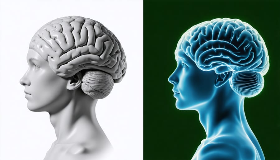
The hippocampus and prefrontal cortex are among the brain regions most affected by Delta-9 THC, as revealed through neuroimaging studies.
The Reward Center Lights Up
When someone uses cannabis, their brain essentially throws a little celebration—and neuroimaging studies let us watch it happen in real time. The star of this show is the mesolimbic pathway, often called the brain’s reward circuit, which runs from the ventral tegmental area to a small structure called the nucleus accumbens.
Think of the nucleus accumbens as your brain’s enthusiasm button. When you eat delicious food, win a game, or receive unexpected good news, this region lights up on brain scans as dopamine floods the area. Delta-9 THC activates the same system, which is why researchers using functional MRI and PET scans consistently observe increased activity in these pleasure centers during cannabis use.
Here’s what makes this fascinating: THC doesn’t just stimulate these regions directly. Instead, it works more like a backstage director, influencing the cannabinoid receptors that regulate dopamine release. When THC binds to CB1 receptors in this pathway, it essentially removes the brakes from dopamine-producing neurons, allowing more of this “feel-good” neurotransmitter to flow freely.
Neuroimaging studies reveal this process beautifully—brain scans show the nucleus accumbens glowing brighter during THC exposure, corresponding directly with users’ reports of euphoria and pleasure. It’s the same neural signature scientists observe with other rewarding experiences, explaining why that feeling of contentment or giggly happiness emerges. Understanding this mechanism helps researchers comprehend not just cannabis’s pleasurable effects, but also its potential for habitual use in some individuals.
Memory and the Foggy Hippocampus
Have you ever walked into a room and completely forgotten why you went there? That momentary mental fog is something we’ve all experienced, and it turns out THC creates a remarkably similar effect in your brain—except neuroimaging lets us watch it happen in real time.
When researchers use fMRI and PET scans to observe cannabis users, the hippocampus—your brain’s memory formation center—shows noticeably altered activity patterns. Think of the hippocampus as your brain’s filing clerk, constantly sorting and storing new information. Under THC’s influence, this clerk seems to take an extended coffee break. The imaging reveals decreased blood flow and reduced communication between the hippocampus and other memory-related regions.
This isn’t just abstract neuroscience—it explains that classic cannabis experience where you start telling a story and midway through, lose your train of thought entirely. One study showed participants a series of words while under THC’s effects. The brain scans revealed their hippocampus struggled to “light up” during the memory encoding process, like trying to save a document on a computer with a glitchy hard drive.
What’s particularly fascinating is that these effects appear temporary for most users. Once THC clears your system, the hippocampus generally returns to its normal bustling activity. However, chronic heavy use, especially when started during adolescence, shows more persistent changes in hippocampal structure and function—a finding that’s prompted serious conversations about the timing and frequency of cannabis use.
When the Prefrontal Cortex Takes a Break
Picture your brain’s CEO—the prefrontal cortex—deciding to take an unexpected vacation. That’s essentially what neuroimaging studies reveal when Delta-9 THC enters the picture. This region, nestled right behind your forehead, normally acts as your brain’s executive manager, juggling decision-making, impulse control, and rational thinking. But when THC binds to cannabinoid receptors, brain scans show something fascinating: activity in this crucial control center dramatically decreases.
Think of it like dimming the lights in a busy office. The workers (your neurons) are still there, but they’re operating in a different mode. This reduced prefrontal activity helps explain why your usually cautious friend suddenly thinks ordering three different pizzas at 2 AM is brilliant, or why time seems to stretch like taffy during a cannabis experience. Without your brain’s CEO calling the shots as vigorously, the filters we normally apply to our thoughts and perceptions relax.
Functional MRI studies have captured this phenomenon beautifully, showing cooler colors—representing less activity—washing over the prefrontal regions. Meanwhile, other brain areas light up more intensely, particularly those involved in sensory processing and emotion. It’s not that your brain stops working; rather, it shifts priorities. The rational planner takes a backseat while the creative dreamer grabs the wheel, fundamentally altering how you process information and make decisions in those moments.
The Sensory Experience Amplified
Have you ever noticed how music seems to wrap around you differently after cannabis use, or how colors appear more vivid? Neuroimaging studies reveal what’s happening behind these sensory shifts. When Delta-9 THC enters your system, brain scans show heightened activity in regions like the primary visual cortex and auditory processing centers—the areas responsible for interpreting what you see and hear.
Think of your brain’s sensory filters as volume knobs that THC turns up. Research using fMRI demonstrates increased connectivity between sensory regions and emotion centers like the amygdala, which might explain why that song suddenly feels profoundly moving or why ordinary textures become fascinating. One study participant described touching velvet as “seeing sound,” illustrating how THC can blur the boundaries between different sensory channels—a phenomenon called synesthesia.
The thalamus, your brain’s sensory relay station, also shows altered activity patterns under THC’s influence. Instead of filtering sensory information as usual, it becomes more permissive, allowing details you’d typically ignore to flood into conscious awareness. This neurological shift explains why mundane experiences—watching clouds drift or tasting food—can transform into captivating events that demand your complete attention.
Acute vs. Chronic: Different Pictures in the Scanner
Your brain is remarkably adaptable—think of it like a seasoned coffee drinker who eventually needs two cups to feel what one used to provide. When it comes to Delta-9 THC, the difference between someone trying cannabis for the first time and someone who’s used it daily for years shows up dramatically in brain scans, revealing a fascinating story of neural adaptation.
When researchers compare acute users (those experiencing their first or occasional exposures) with chronic users, they’re essentially watching two different movies. In acute users, THC lights up the brain like a fireworks display—the cerebellum buzzes with activity affecting coordination, the limbic system responds intensely to emotional stimuli, and the prefrontal cortex shows significant alterations in decision-making regions. It’s overwhelming because the brain hasn’t learned to compensate yet.
Chronic users tell a different story. Their scans often show what scientists call “blunted responses”—the same dose of THC produces less dramatic changes in blood flow and activity patterns. This isn’t necessarily good news; it’s evidence of tolerance. The brain has downregulated its cannabinoid receptors, similar to turning down the volume on speakers that have been blasting too long. Studies using PET scans have found that heavy, long-term users can have up to 20% fewer CB1 receptors in certain brain regions compared to non-users.
But here’s where it gets interesting: some of these changes appear reversible. Research following people after they stop using cannabis shows that receptor density can bounce back, sometimes within weeks, though complete recovery may take months. The brain demonstrates remarkable resilience.
The more concerning findings involve structural changes. Some studies using MRI have reported reduced volume in the hippocampus and amygdala among chronic users—regions critical for memory and emotion. However, other research has failed to replicate these findings, and scientists debate whether observed changes represent actual tissue loss or temporary alterations in how brain cells hold water and nutrients.
What’s particularly intriguing is that not all chronic users show the same patterns. Age of first use matters significantly—adolescent brains appear more vulnerable to lasting changes than adult brains. Frequency and potency also play roles, as does individual genetic variation in how people metabolize THC.
The brain scans essentially reveal a negotiation: acute exposure shows disruption, chronic exposure shows adaptation, but adaptation comes at a cost that may or may not be fully reversible.
The Age Question: Why Young Brains Show Different Patterns
The teenage brain isn’t simply a smaller version of an adult brain—it’s more like a construction site, actively remodeling itself in profound ways. This ongoing renovation project helps explain why neuroimaging studies reveal strikingly different patterns when comparing adolescent and adult responses to Delta-9 THC.
Think of brain development like building a house. During adolescence, the brain is still installing critical wiring, particularly in the prefrontal cortex—the area responsible for decision-making, impulse control, and planning. Simultaneously, the brain is pruning unnecessary connections, strengthening important pathways, and fine-tuning its communication networks through a process called myelination. This construction phase typically continues until the mid-twenties.
When researchers use fMRI to examine adolescent brains exposed to THC, they observe something concerning: the drug appears to interfere with this construction process. Studies from institutions like the University of Vermont have documented differences in how adolescent brains activate reward circuits compared to adults. Young brains show heightened responses in areas like the nucleus accumbens—the brain’s pleasure center—which may partly explain why teenagers can be particularly vulnerable to developing cannabis use patterns.
More intriguingly, longitudinal neuroimaging studies tracking the same individuals over time suggest that regular THC exposure during adolescence correlates with altered trajectories in brain development. Regions still under construction seem particularly susceptible to disruption. One study following participants from ages 14 to 22 found that those who used cannabis heavily during this window showed reduced connectivity in neural networks critical for memory and attention.
However—and this is crucial—these findings don’t translate into simple doom-and-gloom narratives. Many factors influence outcomes, including frequency of use, potency, individual genetics, and environmental influences. The research suggests heightened caution during developmental years, not that any exposure guarantees harm. Understanding these vulnerabilities helps young people make informed choices while their brains complete their remarkable construction project.
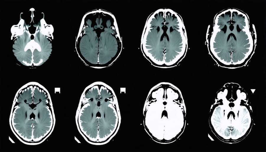
Brain imaging research reveals significant differences in how THC affects adolescent versus adult brains, particularly in areas still undergoing development.
What We Still Don’t Know (And Why That Matters)
Despite decades of research, our understanding of THC’s effects on the brain remains frustratingly incomplete—like trying to solve a puzzle when you’re not even sure you have all the pieces.
One major limitation is that most neuroimaging studies involve relatively short-term observations. We can watch what happens when someone uses cannabis today, but tracking brain changes over years or decades presents enormous logistical challenges. It’s the difference between photographing a tree in different seasons versus watching a forest ecosystem evolve over centuries. Long-term studies require consistent funding, participant retention, and technology that remains compatible across many years—a tall order in our rapidly advancing field.
There’s also the chicken-and-egg problem: When we see differences in brain activity or structure between cannabis users and non-users, what came first? Did cannabis change the brain, or were those brains already different in ways that made cannabis use more likely? Imagine studying whether coffee changes your morning routine—but some people are naturally early risers who happen to enjoy coffee. Teasing apart cause from correlation requires sophisticated study designs that are expensive and time-consuming.
Individual variation presents another puzzle. Why does cannabis affect my cousin’s anxiety so differently than mine? We suspect genetics, prior experiences, and even gut bacteria might play roles, but neuroimaging typically shows us group averages, potentially obscuring crucial individual differences. It’s like understanding “average weather”—technically accurate but not particularly useful for planning your specific Tuesday.
Perhaps most importantly, we’re still refining what “normal” brain function even looks like. Every brain is wonderfully unique, making it challenging to define what constitutes a meaningful change. These gaps aren’t failures of science—they’re invitations to keep asking better questions.
Neuroscientists continue to analyze brain imaging data to understand the full scope of THC’s effects and identify remaining research questions.
When you think about getting high, you’re experiencing a profoundly biological event—not just an abstract state of mind, but actual molecules binding to receptors, blood flow patterns shifting, and neural networks reorganizing their conversation. Neuroimaging has given us something remarkable: the ability to watch these invisible processes unfold in real time, transforming subjective experience into measurable reality.
The brain scans we’ve explored reveal THC as both more subtle and more complex than cultural stereotypes suggest. It doesn’t simply “shut down” the brain or make everything fire randomly. Instead, it recalibrates specific networks—dimming the executive control center while amplifying sensory and memory-processing regions, loosening the default mode network’s grip while potentially strengthening connections between areas that don’t usually chat so freely. These patterns help explain why a cannabis high feels simultaneously relaxing and perceptually vivid, why time seems elastic, and why that song suddenly sounds transcendent.
Perhaps most importantly, neuroimaging reminds us that variation is the rule, not the exception. Your brain’s response to THC reflects your unique biology, history, and context. As research continues, we’re not just mapping what cannabis does to *the* brain—we’re learning what it does to *your* brain, opening doors to more personalized understanding of this ancient plant’s relationship with human consciousness.


Leave a Reply